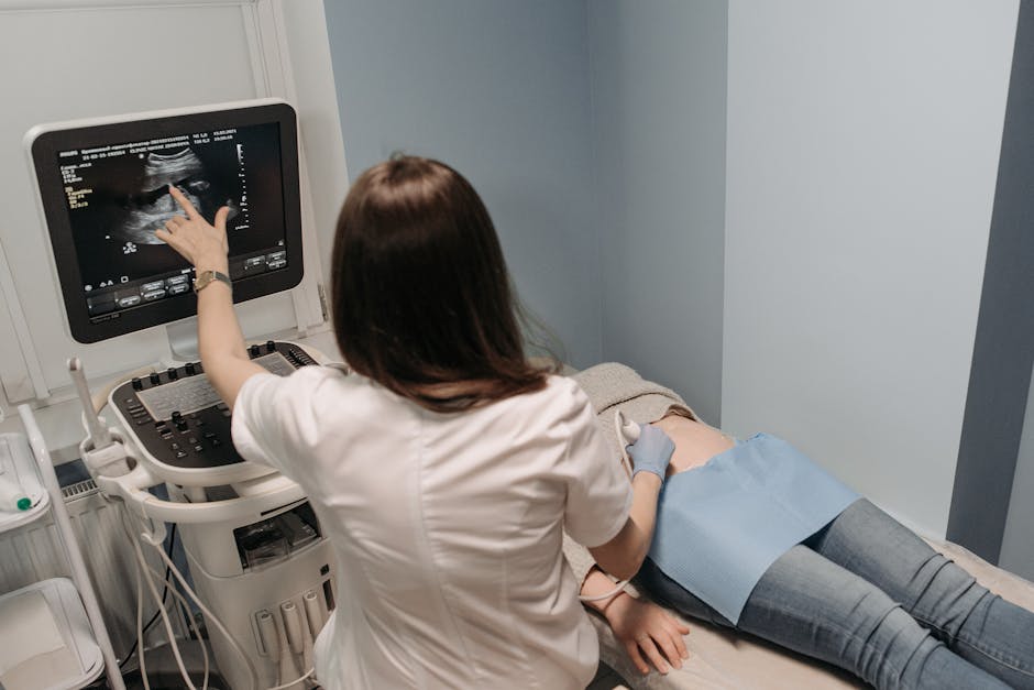
Dr. John Kugler, a seasoned hospitalist at Stanford known for his expertise in point-of-care ultrasound (POCUS), recently shared valuable insights on lung ultrasound in a captivating video presentation. In his practice, lung ultrasound plays a pivotal role in diagnosing various conditions such as pneumothorax, pleural effusions, and distinguishing between A-lines and B-lines to detect pulmonary edema or pneumonia.
When it comes to diagnosing pneumothorax, a critical condition where air accumulates between the lung's layers, Dr. Kugler emphasizes its significance not only in emergency settings but also in hospital medicine, especially post-invasive procedures. He sheds light on the key steps involved in using lung ultrasound to identify pneumothorax:
Firstly, selecting the appropriate probe, ideally a linear one for clearer images, although other probes can suffice. Secondly, positioning the patient flat to aid in air detection within the chest cavity, facilitating easier pneumothorax identification. Lastly, recognizing lung sliding as a normal finding, where the visceral and parietal pleura move synchronously during breathing, akin to "ants on a log," as described by Dr. Kugler.
When it comes to detecting pleural effusions, lung ultrasound stands out for its efficacy, especially in identifying small fluid amounts that might escape detection on chest X-rays. Dr. Kugler recommends using a cardiac probe for this scan and advises patients to position their arm behind their head to expose the chest side adequately, particularly the axillary region where fluid tends to collect.
On the right side, the liver serves as an excellent acoustic window for imaging, while on the left, the smaller and more variable spleen poses challenges. Additionally, scanning the left side can be complicated due to the spleen's size and positioning variability, along with potential interference from a stomach bubble affecting image clarity.
Dr. Kugler highlights key ultrasound signs for pleural effusions, including the "spine sign," a crucial indicator where the spine should not be visible above the diaphragm under normal conditions due to air-filled lungs blocking ultrasound waves. However, the presence of fluid makes the spine visible, signaling a pleural effusion.
In conclusion, lung ultrasound, with its ability to provide real-time imaging and aid in diagnosing various pulmonary conditions, proves to be an invaluable tool in the hands of experienced practitioners like Dr. Kugler. As technology continues to advance, the role of ultrasound in healthcare is only expected to grow, revolutionizing diagnostic practices and improving patient care.