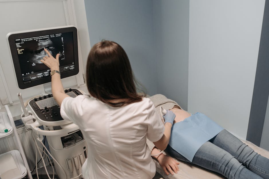
Ultrasound technology has revolutionized the field of medical imaging, allowing for non-invasive visualization of internal structures and abnormalities. In a recent study, researchers sought to investigate the use of Ultrasonographic vascular imaging in identifying superficial invasive lymphomas and indolent lymphomas. This retrospective study enrolled 82 lymphoma patients and utilized three established ultrasound techniques: color Doppler flow imaging (CDFI), angio plus ultrasound imaging (AngioPLUS™), and contrast-enhanced ultrasound (CEUS).
The lymph nodes were classified as invasive or indolent lymphomatous lymph nodes based on proliferation rates and clinical course. The researchers then compared qualitative and quantitative parameters from the two groups to determine the differences in blood flow distribution. The results showed statistically significant differences in the types of blood flow distribution between invasive and indolent lymphomatous lymph nodes across all three ultrasound techniques.
In CDFI, the resistance index (RI) was found to be a determining factor in identifying invasive or indolent lymphomatous lymph nodes. AngioPLUS™ demonstrated better blood flow performance and diagnostic sensitivity compared to CDFI. Meanwhile, CEUS showed statistically significant differences between the two groups in parameters such as necrosis and arrival time (ATM).
Combining CDFI with CEUS resulted in the highest diagnostic sensitivity of 98.1%, offering clinicians a valuable tool for accurately differentiating between invasive and indolent lymphomas. The study also highlighted the excellent interobserver agreements for qualitative parameters, further emphasizing the reliability of Ultrasonographic vascular imaging in lymphoma diagnosis.
According to the World Health Organization Classification of Lymphoid Neoplasms, lymphomas encompass a wide range of subtypes with varying clinical behaviors. The distinction between invasive and indolent lymphomas is crucial for guiding treatment decisions and predicting patient outcomes. In cases of transformation from low-grade indolent NHL to a more aggressive form, accurate identification of the lymphoma type is essential for appropriate management.
The findings of this study underscore the importance of utilizing advanced imaging techniques like Ultrasonographic vascular imaging in lymphoma diagnosis. By combining multiple ultrasound modalities, clinicians can improve their ability to differentiate between different types of lymphomas and tailor treatment strategies accordingly. This research contributes to the growing body of evidence supporting the use of ultrasound in oncology and highlights the potential for further advancements in ultrasound technology for cancer imaging.
As medical imaging continues to evolve, incorporating innovative techniques like contrast-enhanced ultrasound and high-frequency ultrasound in lymphaticovenous anastomosis and lymph node characterization, the field of oncology stands to benefit from improved diagnostic accuracy and patient care. The integration of ultrasound artifact identification in lymphoma diagnosis represents a significant step forward in personalized medicine, offering clinicians a powerful tool for optimizing treatment outcomes and enhancing patient quality of life.