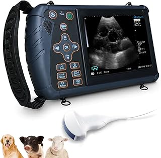
An echocardiogram, commonly referred to as an “echo,” is a non-invasive scan used to examine the heart and surrounding blood vessels. This diagnostic procedure utilizes ultrasound technology, where high-frequency sound waves are emitted by a small probe. These waves create echoes when they interact with different areas of the body, which are then captured by the probe and translated into real-time images displayed on a monitor.
This imaging technique is invaluable in assessing various heart conditions by providing insights into the heart’s structure, blood flow patterns, and the functionality of its pumping chambers. It plays a crucial role in diagnosing issues such as heart attacks, heart failure, congenital heart defects, problems with heart valves, cardiomyopathy, and endocarditis. An echocardiogram is instrumental in guiding healthcare professionals in determining the most appropriate treatment strategies for these cardiac conditions.
An echocardiogram is typically requested by a cardiologist or any physician suspecting heart-related problems in a patient. The procedure is commonly performed in a clinical setting by trained professionals like cardiologists, cardiac physiologists, or sonographers. It is essential to note the distinction between an echocardiogram and an electrocardiogram (ECG), with the latter primarily focusing on assessing heart rhythm and electrical activity.
The echocardiogram procedure involves different methods, with the transthoracic echocardiogram (TTE) being the most common approach. During a TTE, the patient lies on a bed after removing clothing from the upper body. Electrodes are attached to the chest to monitor heart rhythm, and a gel is applied to aid in ultrasound probe movement. The probe is then maneuvered across the chest, capturing images that are displayed and recorded by a nearby machine. The entire process typically lasts between 15 to 60 minutes.
Aside from the TTE, there are several other types of echocardiograms available for specific diagnostic purposes. These include the transoesophageal echocardiogram (TOE), stress echocardiogram, and contrast echocardiogram. Each variant caters to different clinical scenarios, providing detailed imaging based on the heart condition being evaluated. For instance, a stress echocardiogram may be recommended for individuals whose heart issues manifest during physical exertion.
After undergoing an echocardiogram, the results are usually interpreted by healthcare professionals before being communicated to the patient during a follow-up appointment. The procedure is generally safe and painless, with no radiation involved in standard echocardiograms. However, some variants like the TOE may cause temporary discomfort, and there are potential risks associated with procedures involving sedation or contrast agents.
In conclusion, echocardiography serves as a vital tool in the diagnosis and management of various cardiac conditions. Its non-invasive nature, coupled with the ability to provide detailed real-time images of the heart, makes it a cornerstone in modern cardiology practice, aiding in delivering optimal patient care and treatment outcomes.
📰 Related Articles
- e.l.f. Beauty & Pinterest Launch AI Color Analysis Tool
- Wedding Websites: The Modern Essential for Couples and Guests
- Wandering Spleen in Pediatric Patients: Diagnostic Challenges and Management
- WWII Veteran’s Purple Heart Returned Home by Nonprofit Group
- Vietnam War Veteran Receives Long-Awaited Purple Heart Recognition


