A trusted destination for sonography and ultrasound education resources, diagnostic imaging guides, scanning references, anatomy learning tools, and professional materials for students and healthcare practitioners.
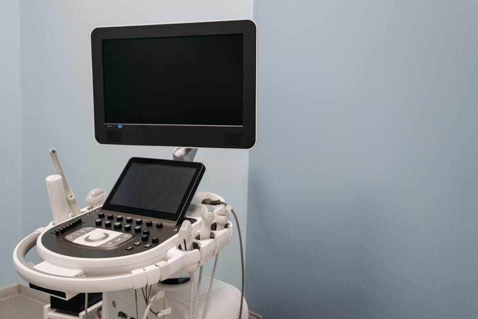
Abdominal ultrasound protocols are integral to the field of diagnostic imaging, providing valuable insights into the internal structures of the abdomen. These protocols guide healt
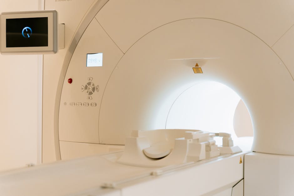
The demand for cardiovascular ultrasound in the USA is on the rise, with projections indicating a significant increase from USD 0.5 billion in 2025 to USD 1.0 billion by 2035. This
Radiologists play a crucial role in diagnosing and treating patients, spending long hours analyzing images on computer screens. The rise in workload and extended screen time has le
The intricate process of prenatal brain development can be significantly influenced by various factors, including stress and exposure to environmental pollutants. Recent research h
In the ever-changing world of healthcare, advancements in technology play a pivotal role in driving progress. From improving patient outcomes to streamlining processes, innovations
As a nurse, you possess a unique set of skills that are highly transferable to a variety of professions within the healthcare industry. Whether you're looking for a change of pace,